Scientific Program

Conference Series Ltd invites all the participants across the globe to attend 28th World Congress on Neurology & Neuroscience Mövenpick Hotel Zürich Regensdorf, Zurich Switzerland.

Day :

  • Speaker Session
Location: St. Gallen II

Session Introduction

Jennifer Brown

Hillfield Pediatric Dentistry, USA

Title: The components of the adolescent brain and its unique sensitivity to sexually explicit material

Time : 11:40-12:10

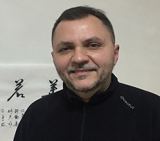
Speaker
Biography:

Dr. Jennifer Brown received her bachelor’s degree in microbiology from Weber State University. She then attended Creighton University Dental School where she received her Doctorate of Dental Surgery. She is the proud mother of five boys.

Abstract:

Introduction: The focus of this brief literature review is to explore whether there is a relationship between the unique anatomical and physiological paradigms of the adolescent brain and an increased sensitivity to sexually
explicit material.
Methods: The EBSCO Research Data bases were searched using the following key terms: adolescence, adolescent brain development, neuroplasticity, sexually explicit material, sexualization, and pornography.
Results: The literature highlighted several components of the adolescent brain that are different than the mature brain. These include: an immature prefrontal cortex and over-responsive limbic and striatal circuits, heightened period for neuroplasticity, overactive dopamine system, a pronounced HPA axis, augmented levels of testosterone, and the unique impact of steroid hormones. The physiological response to sexually explicit material is delineated. The overlap of key areas associated with the unique adolescent brain development and sexually explicit material is noteworthy. A working model summary that compares the response of the adult and adolescent brain to the same sexually explicit stimulus is outlined.
Conclusions: The literature suggests that the adolescent brain may indeed be more sensitive to sexually explicit material, but due to a lack of empirical studies this question cannot be answered definitively. Suggestions for future
research are given to further advance the work in this applicable field of today.

Speaker
Biography:

Dr. Wilma Delphine Silvia CR, MBBS, MD, DNB, MNAMS, has 24 years of teaching experience. She is currently Professor and Head, Department of Biochemistry at Akash Institute of Medical Sciences and Research Centre, Bangalore. She pursued MBBS from Bangalore Medical College, Bangalore and MD from Kasturba Medical College, Manipal. She is the recipient of Excellence award and Basavashree award. Executive Editor of IJCDR and Associate Editor of IJPBS. She has published more than 80 Scientific papers in an International and National Journals and published books on “Mnemonics in Biochemistry” and “PracticalBiochemistry for MBBS Students”. Also a PhD guide, NABL Assessor, NABH Entry Level Assessor and a subject expert for entrance book project under Macula Health Care Pvt Ltd. She has travelled in UK, USA and Europe.

Abstract:

Stress has become part of students’ academic life. Adolescents are particularly vulnerable to the problems associated with academic stress as transitions occur at an individual and social level. Albeit, it becomes imperative to understand the academic stress in order to derive efficient intervention strategies. Hence, the study was aimed for
“Evaluation of stress among medical students during examination using Artificial Intelligence based Graphology and its correlation with salivary cortisol”. This study employed a quantitative research design where 43 medical students (19 males and 24 females) enrolled as subjects, within the age group of 18-26 years. Subjects were monitored to follow the protocol prior to the collection of salivary samples. Salivary samples were collected during pre examination and post examination for cortisol estimation by Competitive ELISA method. Cortisol the “stress hormone” spike during
times of high stress in the body. Salivary cortisol has been used as a biomarker of psychosocial stress and can be indirectly used to assess psychobiological mechanisms that trigger the hypothalamus-pituitary-adrenal axis. Written examination manuscript images were captured for Artificial Intelligence based Graphology analysis. Graphology can be a useful tool in spotting health problems before they become too severe, and is excellent at identifying stress in the individual. The negative traits, which reflects the stress was determined by handwriting were explored in this study. Artificial intelligence (AI) aims to mimic human cognitive functions. In medicine, applications of artificial intelligence have been innovative. The diverse areas of AI include: neural networks, programming languages, genetic algorithms, speech/handwriting recognition etc. The results of the study indicated statistically significant elevated salivary cortisol levels during examination. Students with raised salivary cortisol during post examination levels showed raised negative graphology traits score & low academic performance (P< 0.05). Handwriting was processed through trained Convolutional Neural Network Model of AI. Compared the Negative traits extracted by CNN with
Negative traits determined by Graphology. This study is the first of its kind in India to prove that empirically AI based graphology can be used to assess the stress levels as a screening assistive tool for Psychologist, counselors & academic mentors. It would facilitate the development of effective counselling modules and intervention strategies
in order to help the students to alleviate stress.

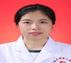
Yuhong Man

The Second Hospital of Jilin University, China

Title: Vitamin B12 , homocysteine level and vascular dementia

Time : 12:40-13:10
Speaker
Biography:

Yuhong Man has completed her PhD at the age of 35 years from Jilin University. She is the group leader of Teaching Group of Behavioral Medicine in Jilin University. She has published more than 35 papers in Chinese journals.

Abstract:

Vitamin B12 , homocysteine level and vascular dementia: This study aim to investigate the relationship of vitamin B12, homocysteine level and vascular dementia. This was a retrospective study and we reviewed 162 patients with brain magnetic resonance imaging (MRI) and mini-mental state examination (MMSE) confirmed admitted patients with vascular dementia (VaD). Vitamin B12 and homocysteine level were assessed to determine their values for predicting functional outcome at the admission first and the follow-up 6 months clinic visits after discharge from the hospital. Associations between vitamin B12, homocysteine level and severity of VaD at admission was analyzed using logistic regression. Results have shown that serum vitamin B12 levels were significantly lower, but the plasma homocysteine level was higher in patients with VaD. High homocysteine levels were independently associated with a decreased risk of MMSE at admission score of VaD (OR 2.87, 95% CI 1.13, 4.63). We found that lower levels of vitamin B12 were associated with worse prognosis at admission and the follow-up 6 months with VaD(OR 3.40, 95% CI 1.16,8.33). Our findings suggested that higher homocysteine levels and lower levels of vitamin B12 was associated with better outcome at admission and the follow-up 6 months with VaD.

Speaker
Biography:

Joao Alexandre Lobo Marques is associate professor at University of Saint Joseph, Macau, SAR China and visiting associate professor at the Chinese Academy of Sciences, Shenzhen (SIAT/CAS), China. He received post doctorate and honorary research fellow from the University of Leicester, UK. He did PhD in Engineering of Teleinformatics at UFC/Brazil and Technische Universität München, Germany (2010). He is the Board Member of the XS Innovation Group in Brazil and former associate professor. He is research and innovation Director at University Lusiada of Angola (2009-2016). He is also associate professor and chief of the software major area at the University UGS, Angola (2016). He has experience in signal processing, applied computer science and artificial intelligence system modeling and implementation, based on several machine learning techniques, artificial neural networks (ANN) and deep learning architectures.

Abstract:

The application of low cost and user friendly equipments must be considered during neuromarketing experiments and projects. The adoption of single channel EEG based on pre frontal cortex monitoring, might provide long and short term information about relevant information for neuromarketing applications, such as attention, focus and relaxation levels. Many studies have been performed using EEG or fMRI for neuromarketing, nevertheless, nonlinear metrics has not been applied yet in a wide and comparative approach. The main goal of this research is to perform nonlinear analysis of attention and relaxation time series during emotional based activities using entropy measures such as Approximate Entropy (ApEn) and Sample Entropy (SampEn), which are template based widely used to measure physiological time series complexity. The considered database consists in 100 experiments from 10 individuals (50% male), ages 24.3 ± 2.00. Three categories of web video advertisements were considered: emotional related, sports relate and food related, with a minimum duration of 45 seconds. Attention and Relaxation levels from the prefrontal cortex were recorded minimum 1 minute before, during and 1 minute after the web video ads. The preliminary results presents that the entropies for attention level changed significantly for the three groups. The entropies for the relaxation level showed significant difference for the emotional related ad category. No significant difference has been stablished for gender analysis. The results are promising and the application of the entropies of attention and relaxation time series as a measure of effectiveness of web video advertisement can be considered. In future studies, a larger number of subjects and a broader range of advertisements are going to be considered.

Speaker
Biography:

Introduction: Primary osteosarcoma (OS) is the second most common primary bone malignancy, the first being multiple myeloma. OS occurs in the second decade, with a predilection for ends of long bones.Head and neck involvement is seen in 2–9% with extragnathic craniofacial bones in 1–2% of cases.Small cell OS (SCO) constitutes 1.3–4% of all OS, skeletal distribution and age range being similar.
Materials–Methods: We report two rares osteosarcoma and we done the review of the litterature about the management and the outcome about intracranial osteosarcoma in our department of neurosurgery.
Results: it is two osteosarcoma cases about a 72-year-old man and one 49-year-old man who both mimiking first meningioma. The first cases is an unsual site parietal and the second case is occipital.the both benefited surgery with excision and exam of histology confirm diagnosis. But the first case died 15 days after surgery in intensive unit care and the second cases died after one year, he benefited surgery and chemotherapy.
Conclusion: Small cell osteosarcoma (SCO) is an extremely uncommon entity that mainly involves the metaphysis of long bones and, rarely, the skull. Histopathology is the key to establishing the correct diagnosis, including subtyping for appropriate management and prognostication, as radiological features are not specific.

Abstract:

BANKOLE Nourou Dine Adeniran born on 11/03/1991 in Cotonou / BENIN, Single. He started medical studies in 2009 and he had been graduated in 2016 Doctor in medicine at Senegal. He is a great passionate of neuroscience including Neurosurgery. Now, he is in 3rd year of residency training in neurosurgery at the Faculty of Medicine and Pharmacy of Rabat / Mohamed V University, WFNS reference center for training of African Neurosurgeons. He is the principal author of this publication in literature 2018 “Child Spinal Cord Compression without Trauma, 113 Cases in Senegal. Yangtze Medicine, 2, 114-128.’’

Speaker
Biography:

Abstract:

Introduction: Primary brain tumors, including brainstem gliomas, are a diverse group of diseases that together constitute the most common solid tumor of childhood. Immunohistochemical analysis, cytogenetic and molecular genetics findings, and measures of mitotic activity are increasingly used in tumor diagnosis and classification. Most children with DIPG are diagnosed between the ages of 5 and 10 years. Focal pilocytic astrocytomas in the brain stem occur less frequently. The goals of our study is to highlight through a literature review since the 1975s to nowadays the progress in the management of DIPG in children and to reach a consensus in our contexts in Africa and to share our experience with multimodal management results about 12 cases in our neurosurgery department.
Materials –Methods: This is a retrospective monocentric study (Department of Neurosurgery Specialty Hospital of Rabat). Included are all children with DIPG Brainstem who managed between January 2008 to December 2018. We studied sex, age, management.
Results: 12 patients were included. The Average age is 6.4Y with extremis 1.3year to 17Y. The sex ratio is two with 67% of boys and 13% of girls. We performed BST on three cases to have histology, and two patients have focal resection, 6 patients benefited shunt (VCS, DVP), and all of our patients had benefited neurooncology treatment, and we have three cases who are benefited directly chemotherapy and X-Ray Therapy before multidisciplinary staff and didn’t benefited any surgery procedure.
Conclusion: Brain tumors have classified according to histology, but tumor location and extent of spread are important factors that affect treatment and prognosis. Approximately 300 to 400 pediatric brain stem tumors are diagnosed each year in the United States. DIPG accounts for approximately 75% to 80% of pediatric brain stem  tumors and constitue 15-20% of all CNS tumours in children and the main cause of death.